Accurate diagnosis is the first step toward effective treatment. Our orthopedic specialists use advanced diagnostic imaging to evaluate injuries and conditions affecting the bones, joints, and soft tissues. These tools allow our team to identify the exact cause of pain, track healing progress, and plan surgical or nonsurgical treatment with precision.
Common Imaging Services We Use:
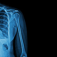
- X-rays (Radiographs): The most common imaging tool in orthopedics, X-rays help visualize bones and joint alignment, detect fractures, arthritis, and other bone abnormalities.
- Ultrasound: Used to assess muscles, tendons, and ligaments in real time. Ultrasound is especially helpful for guiding joint injections and evaluating soft tissue injuries.
- MRI (Magnetic Resonance Imaging): Provides highly detailed images of soft tissues, including ligaments, cartilage, and tendons—often used to diagnose tears or other internal joint problems.
- CT (Computed Tomography) Scans: Offers cross-sectional images of the body and is particularly useful for evaluating complex fractures or planning for reconstructive surgery.
- Bone Scans (when indicated): Detects areas of increased bone activity, which may indicate infection, stress fractures, or certain bone diseases.
Why Imaging Matters
These diagnostic tools allow our orthopedic team to:
- Confirm or rule out specific conditions
- Guide treatment decisions and surgical planning
- Monitor healing and post-surgical recovery
- The imaging services performed at our office include X-rays and Ultrasounds.