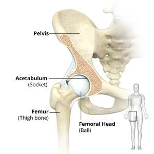
Hip Anatomy
The hip joint is the largest weight-bearing joint in the human body. It is also referred to as a ball and socket joint and is surrounded by muscles, ligaments, and tendons. The thigh bone or femur and the pelvis join to form the hip joint.
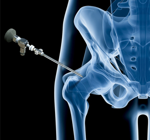
Ultrasound Guided Hip Injections
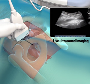
An ultrasound scan is an imaging procedure that uses high-frequency sound waves to produce pictures of the inside of the body. Ultrasound-guided hip joint injections are used to diagnose the underlying cause and relieve hip pain.
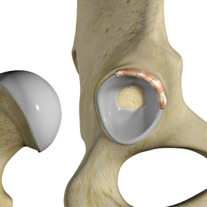
Hip Cartilage Repair
Hip cartilage is a white, tough, flexible tissue covering the ball (femoral head) and socket (acetabulum) of your hip joint. It acts as a cushion or shock-absorber and allows the bones to slide over one another by providing a smooth surface in the joint.
Piriformis Tendon Release

Piriformis tendon release is a surgical procedure that involves the excision or cutting of the piriformis tendon in the hip to reduce pain and improve range of motion.
Hip Labral Repair
Hip labral repair is a surgical procedure to treat hip labral tears. A hip labral tear is a partial or complete rupture of the hip labrum, a ring of fibrous cartilaginous tissue that surrounds the socket of the hip joint.
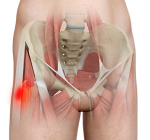
Proximal Hamstring Repair
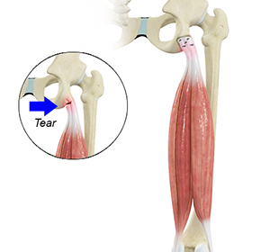
Proximal hamstring injuries can usually be treated with non-surgical options such as the RICE protocol, immobilization, and physical therapy.
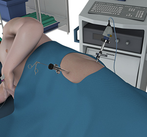
Hip Arthroscopy
Hip arthroscopy, also referred to as keyhole or minimally invasive surgery, is a procedure in which an arthroscope is inserted into your hip joint to check for any damage and repair it simultaneously.
Postless Hip Arthroscopy
Postless hip arthroscopy is a minimally-invasive surgical technique that does not involve the use of perineal posts. Surgery is performed by arthroscopy. An arthroscope is a small, fiber-optic instrument consisting of a lens, light source, and video camera.
Gluteal Strain
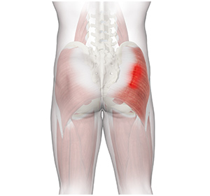
A gluteal strain is a condition characterized by a partial or complete tear of the gluteus muscles, also known as the buttocks. The gluteus muscles are a group of strong muscles present at the back of the pelvis.
Hip Injury
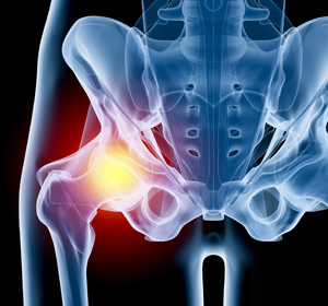
A hip fracture is a break that occurs near the hip in the upper part of the femur or thighbone. It is most frequently caused after minor trauma in elderly patients, and by a high-energy trauma or serious injury in young people.
Gluteus Tendon Tear
The gluteal muscles (situated in the buttocks) are necessary for the stability and movement of the hip joints. The tendons of two gluteal muscles (gluteus medius and gluteal minimus) are attached at the outer hip region and are often called the “rotator cuff of the hip.”
Snapping Hip Syndrome
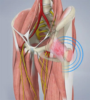
Snapping hip syndrome is a condition in which you hear or feel a snapping sound in the hip when you swing your legs, run, walk or get up from a chair. The sound can be experienced in the back, front or side of the hip.
Hip Bursitis
Hip bursitis is a painful condition caused by the inflammation of a bursa in the hip. Bursae are fluid-filled sacs present in the joints between bone and soft tissue to reduce friction and provide cushioning during movement.
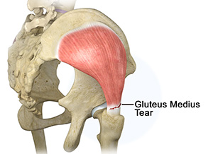
Gluteus Medius Tear
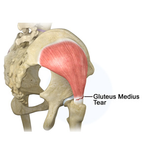
A gluteus medius tear is the partial or complete rupture of the gluteus medius muscle due to severe muscle strain. Gluteus medius tears often occur at the tendinous attachment to the greater trochanter of the femur bone.
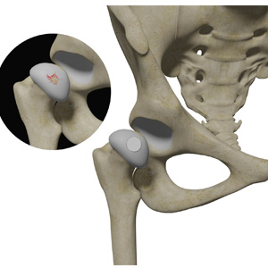
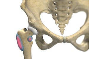
Hip Labral Tear
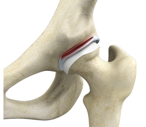
A hip labral tear is an injury to the labrum, the cartilage that surrounds the outside rim of your hip joint socket.
Hip Instability
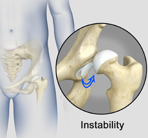
Injury or damage to these structures can lead to a condition called hip instability when the joint becomes unstable.
Hip Synovitis
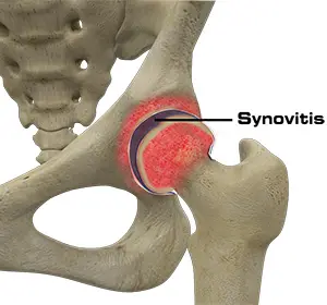
Hip synovitis, also called transient hip synovitis or toxic synovitis, is a condition characterized by inflammation of the synovial tissues that surround the hip joint.
Hip Tendonitis
Tendons are strong connective tissue structures that connect muscle to bone. Hip tendonitis is a condition associated with degeneration of the hip tendons.
Proximal Femoral Fracture
A proximal femoral fracture also known as a hip fracture is a condition characterized by a break or fracture in the upper part of the femur or thighbone in close proximity to the hip socket.
Hamstring Injuries
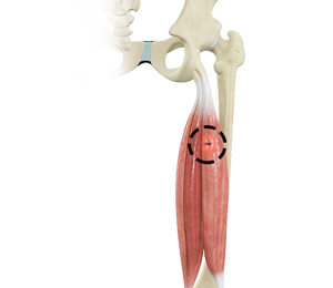
The hamstring is a group of three muscles that run along the back of the thigh from the hip to the knee. Hamstring injuries occur when these muscles are strained or pulled.